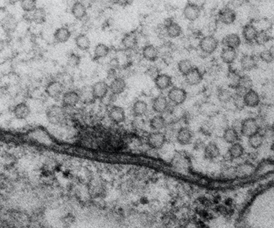
Individual synapse visualized by electron microscopy.
Credit: Serpe Lab, NICHD
Transmission of chemical signals through synapses—structures that connect nerve cells to other neurons and non-neuronal cells—is essential for information processing in the nervous system. Synapses develop and mature via tightly regulated processes that are closely linked to synapse activity, but the mechanisms by which neurons sense synaptic activity are poorly understood.
The Serpe Lab is examining how a particular class of developmental signals, the bone morphogenetic proteins (BMPs), influences the formation of synapses and optimizes their function. In several studies using the fruit fly neuromuscular junction (NMJ) as a model, they discovered a novel, local BMP signaling pathway and demonstrated that it serves as a sensor for synapse activity.
The lab discovered that a molecule called Neto is essential for synapse development. In the absence of Neto, NMJ synapses do not form, and fly embryos die paralyzed. The scientists found that Neto is an obligatory component of the glutamate-receptor complexes required for the assembly of functional NMJs. When they examined BMP signaling in flies with disrupted Neto activity, they found that the effector of the BMP signaling pathway, a transcription factor, accumulates at synaptic terminals in response to synaptic activity.
Recently, Dr. Serpe and her colleague reviewed the main features of this sensor and discussed its function beyond monitoring synapse activity. They described how local BMP signaling serves as a key controller that coordinates with other BMP signaling pathways to balance synapse growth, maturation, and function.
Learn more about the Cell Regulation and Development Affinity Group: https://www.nichd.nih.gov/about/org/dir/affinity-groups/CRD
 BACK TO TOP