Home
We strive to understand fundamental relationships between function and structure in living tissues, primarily in neural tissue and in extracellular matrix (ECM). Specifically, we are interested in how microstructure, hierarchical organization, composition, and material properties all affect biological function and dysfunction. We investigate biological and physical model systems, such as “engineered” tissue constructs and tissue analogs at different time and length scales. These physical measurements are guided by mathematical and computational models we develop to 1) aid in the design of experiments, 2) interpret their findings, 3) make new predictions and 4) generate novel hypotheses. Primarily, we use water molecules to probe both equilibrium and dynamic interactions among tissue constituents over a wide range of time and length scales. At macroscopic length scales we vary water content or ionic composition to determine the equilibrium osmo-mechanical properties of well-defined model systems. To probe tissue microstructure and microdynamics, we employ atomic force microscopy (AFM), small-angle X-ray scattering (SAXS), small-angle neutron scattering (SANS), static light scattering (SLS), dynamic light scattering (DLS), and one-dimensional and multi-dimensional nuclear magnetic resonance (NMR) relaxometry, diffusometry and exchange. To better understand structure/function relationships, we develop and use physics and engineering principles to describe how observed changes in tissue properties, microstructure and composition affect physiological processes by characterizing the transport of mass, charge, momentum, energy, and magnetization. The latter provides the most direct and noninvasive means for describing essential transport processes in vivo. Not surprisingly, we use magnetic resonance imaging (MRI) transport measurements as a vehicle to translate novel quantitative methodologies we develop, and the understanding we glean from them, from "bench to bedside."
Our “bottom up” tissue sciences activities described above dovetail with our basic, applied and translational research in quantitative imaging, which uses a “top down” approach to generate in vivo measurements and maps of intrinsic physical quantities, such as magnetization, diffusivity, relaxivity, and exchange rates, rather than qualitative images widely used in radiology. Our quantitative imaging group uses knowledge of physics, engineering, applied mathematics, imaging sciences, and computer sciences, as well as insights gleaned from our tissue sciences research, to discover and develop novel imaging data derived "stains" and "contrasts" that can sensitively and specifically detect changes in tissue composition, microstructure, or microdynamics. Our goal is to use these new parameters as quantitative imaging biomarkers to assess normal and abnormal development, diagnose childhood diseases and disorders, and characterize degeneration and trauma. MRI is our in vivo imaging method of choice because it is so well-suited to many NICHD mission-critical applications—it is non-invasive, non-ionizing, generally requires no exogenous contrast agents or dyes, and is deemed safe for use with pregnant mothers and their developing fetuses, as well as with children in both clinical and research settings. One technical objective of ours has been to transform clinical MRI scanners into quantitative scientific instruments capable of producing reproducible, accurate, and precise imaging data with which to measure and map useful imaging biomarkers for pre-clinical and clinical applications, including single scans, longitudinal and multi-center studies, personalized or point-of-care medicine, and for populating curated imaging databases.
SQITS News
NIH Lays Groundwork for Advances in Brain Imaging (NIH Record, January 2, 2026)
Neuroscience Grand Rounds: Microstructure and Transport Processes in the Brain with MRI (December 9, 2025)
Basser Experiences Eureka Moment at NIH Research Festival (NIH Record, November 8, 2024)
DIR Showcase: Unraveling the Effects of a Noninvasive Brain Cancer Therapy (July 11, 2024)
Dr. Cai and Dr. Basser's paper, Disentangling the Effects of Restriction and Exchange With Diffusion Exchange Spectroscopy , has been selected for the best paper award in the Medical Physics and Imaging section of Frontiers in Physics. (December 18, 2023)
DIR Showcase: Noninvasively Measuring Brain Metabolic Activity (September 8, 2023)
Spotlight: Advancing neuroscience research for children around the world (September 20, 2022)
Press Release: NIH-developed multidimensional MRI can detect “invisible” brain injuries, studies suggest (August 24, 2022)
Dr. Basser gives the Keynote Lecture at the 37th Annual MD-PhD Conference sponsored by the Anschutz Medical School, University of Colorado . Other notable keynote speakers at this conference were Nobel Laureate Craig Mello, Karl Diesseroth, Catherine Bollard, and NIA’s own Jasmine Belkaid.
SQITS is involved in an important research study being sponsored jointly by the Bill and Melinda Gates Foundation (BMGF) and the NICHD to improve the understanding of neuroanatomical trajectories in children worldwide, but particularly in Lower and Middle Income Countries (LMIC).
More about this study
SQITS is involved in an important research study being sponsored jointly by the Bill and Melinda Gates Foundation (BMGF) and the NICHD to improve the understanding of neuroanatomical trajectories in children worldwide, but particularly in Lower and Middle Income Countries (LMIC). A key component of this brain imaging research initiative has been to establish a pediatric neuroimaging consortium intended to deploy portable, low-cost Hyperfine MRI scanners to enable data collection in areas where there is no neuroradiological infrastructure. Aspects of this unique project were the subject of a recent joint NICHD/BMGF-sponsored workshop https://www.nichd.nih.gov/about/meetings/2022/041822
Since about 2005, SQITS lab members, Ferenc Horkay, Carlo Pierpaoli and Peter Basser, began developing and vetting a novel diffusion MRI “phantom” to help calibrate and standardize single-subject, longitudinal, and multi-site human studies to ensure quantitative MRI data could be collected and meaningfully analyzed, compared and/or pooled. A press release when their patent issued https://www.nichd.nih.gov/newsroom/releases/051917-basser describes a stable, safe, and well-characterized polymer, polyvinylpyrrolidone (PVP), having appropriate tissue-like diffusion properties, which are well-suited to this application.
Fast forward to 2022, the BMGF/NICHD neuroimaging consortium will now be adopting the CaliberMRI phantom provided by QMRI https://qmri.com/ , which incorporates SQITS’ PVP polymer phantom material to standardize diffusion MRI data acquired on different Hyperfine MRI scanners in LMIC sites as well as in MRI physics labs, where other pediatric neuroimaging consortium scientists are developing new MRI methods to advance this important project.
Another related development is that the National Institute of Standards and Technology (NIST) has been testing and promulgating SQITS’ PVP phantom material as an NIST MRI “standard” https://www.nist.gov/news-events/news/2022/05/measuring-phantom-library. According to Stephen Russek, Ph.D., Project Leader for Imaging Physics, NIST in Boulder, CO, who directs this activity, “NICHD should be prominently credited for identifying, developing, and patenting PVP solutions as a diffusion reference. It is remarkably stable and has nearly ideal Gaussian diffusion, which is a rarity among any tissue mimics. As we work to characterize more complex tissue mimics for medical imaging reference standards, we increasingly appreciate PVP solutions.”
Dr. Basser featured as discussant for the Imaging the Future of In vivo Neuropathological Diagnosis through Postmortem Analyses workshop, sponsored by the National Institute of Neurological Disorders and Stroke (NINDS), NIH, July 13-14, 2022.
Elucidating Brain Structure and Architecture (NIH Catalyst, Volume 30, Issue 4, July-August 2022)
VIDEO: Historical ISMRM Campfire (featured at 0:47:00 mark)
VIDEO: Dr. Basser gives the Phil Chen Lecture on Friday, April 29.
Philip Chen Lecture: Using Water Migration to Probe Brain Structure and Architecture
Dr. Basser gives the 2022 Doc Morris Public Lecture on Friday, April 22.
VIDEO: The Senate Committee Reviews Posture, 2023 Authorization Request, Part 1, highlighting important recent work SQITS staff has done (featured at 1:00:00 mark) with our CNRM Neuroradiology – Neuropathology (“Rad-Path”) Core, in collaboration with Dr. Daniel Perl, a USUHS neuropathologist. (April 5, 2022)
VIDEO: International Society of magnetic resonance and medicine (ISMRM) 2020 Lauterbur Lecturer . (August 8-14, 2020)
SQITS partners with the Uniformed Services University's Neuropathology core to form the Neuropathology-Neuroradiology Integration Core.
Dr. Basser receives the Honorary Member Award at the American Society of Neuroradiology (ASNR) 57th Annual Meeting (May 21, 2019)
Carnegie Mellon University College of Engineering News: "Teddy Cai Wins Prestigious Scholarship" (March 29, 2019)
Zilkha Seminar Series, Keck School of Medicine of USC: “Probing Tissue Structure and Dynamics using MRI” (March 13, 2019)
VIDEO (George Mason University Bioengineering Seminar Series): “What can we learn about nervous system structure and function using porous media MR?” (February 28, 2019)
Article on study conducted in collaboration with the Fields lab: Astrocytes Regulate Signal Speeds of Neurons. (Neuroscience News, October 29, 2018)
Press Release: Neurons absorb and release water when firing, NIH study suggests. (September 13, 2018)
VIDEO (NIH Videocast): NICHD Advisory Council Meeting - September 2018 (Dr. Basser is featured at 2:39:19)
Drs. Basser and Tasaki featured in: Fox, D. (2018). Brain Cells Communicate with Mechanical Pulses, Not Electric Signals. Scientific American, (Volume 318, Issue 4), pp.61-67.
BRAIN Initiative: Bridging Gaps in Neuro Knowledge (NIH Catalyst, Volume 25 Issue 6, November–December 2017)
VIDEO (NIH Videocast): "Measuring the latency connectome" (34:00) – Plenary talk, 2017 NIH Research Festival (September 13, 2017)
Medical imaging device developed by NIH researchers awarded U.S. patent (May 19, 2017)
VIDEO (YouTube): "The Invention and Development of Diffusion Tensor NMR and MRI at the NIH" , recorded at a conference hosted by Cardiff University Brain Research Imaging Centre (CUBRIC), January 31 – February 1, 2017
Dr. Derek Jones, Former NIH Mentee, Directs Brain Imaging Center in Wales (NIH Record, July 1, 2016)
VIDEO (YouTube): Dr. Basser featured in "The Human Placenta Project" (more information about the Human Placenta Project)
Johns Hopkins University webcast: “Characterizing brain microstructure, architecture and organization with diffusion MRI” (February 4, 2014)
VIDEO (YouTube): Dr. Basser featured in "Research for a Lifetime: The Journey Forward" to commemorate NICHD's 50th anniversary
Trainee Chinedu Anyaeji selected to participate in the NICHD Scholars Program (The Catalyst, Volume 19, Issue 6, November-December 2011)
"Twitchy Nerves (Literally) May Explain Epilepsy, Pain" on NPR's Morning Edition (October 5, 2010)
Revealing the uncharted brain: frontiers in diffusion medical-resonance imaging (SPIE, March 3, 2009)
Awards
Collaborator Rhea Carlson is the 2025 Bruker MRI Award 3rd place winner at the 2025 ISMRM conference in Hawai'i.
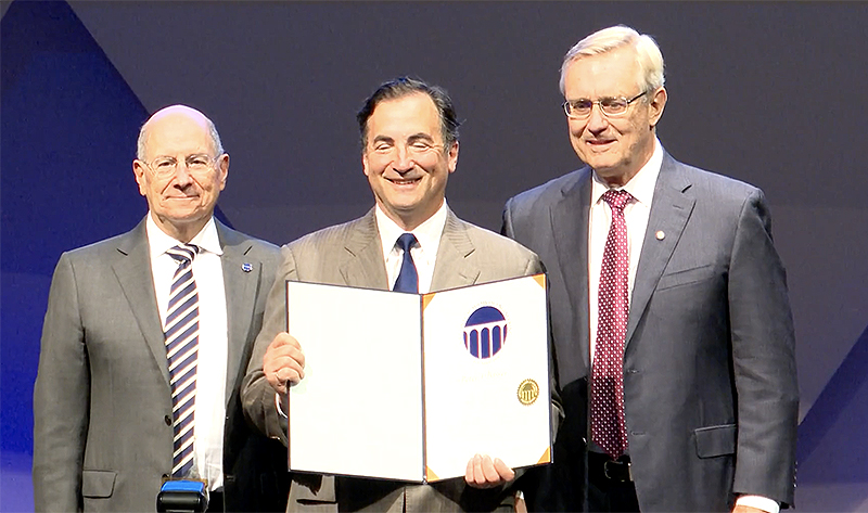
Dr. Basser is inducted into the National Academy of Inventors (NAI) 2024 Class of Fellows. NAI Fellowship is the highest professional distinction awarded solely to inventors. Check out the full list of 2024 Fellows (PDF 438 KB) (December 10, 2024)
VIDEO: Dr. Basser is formally inducted into the Class of 2020 National Academy of Engineering (NAE) , October 2-3, 2022 (featured at 33:30 mark).
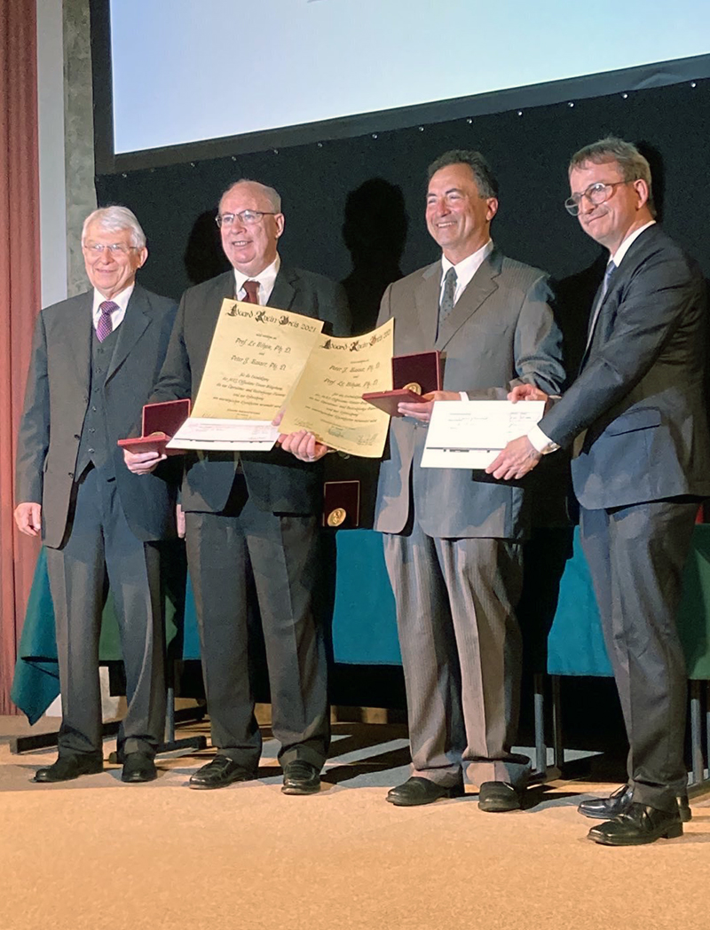
Dr. Basser accepts the Eduard Rhein Technology Award in Munich.
NICHD’s Basser Honored for Developing Imaging Techniques
Dr. Peter Basser is awarded the Technology Award 2021 of the EDUARD RHEIN FOUNDATION.
Dr. Velencia Witherspoon announced as part of Inaugural Cohort of NIGMS MOSAIC Scholars.
Dr. Velencia Witherspoon received a Fellows Recruitment Incentive Award (FRIA) renewal for 2020.
Dr. Peter Basser is inducted to the National Academy of Engineering, 2020 "For development of diffusion tensor MRI and streamline tractography, transforming the characterization of brain disorders and visualization of nerve fiber pathways."
Dr. Peter Basser is awarded the 2019 Honorary Member Award by the American Society of Neuroradiology
From the Deputy Director of Intramural Research's website (screencapture):
Peter Basser, Ph.D., head of the NICHD Section on Quantitative Imaging and Tissue Sciences, has been made an honorary member of the American Society of Neuroradiology, an organization of more than 5,600 neuroradiologists and related professionals.
Basser is a scientist-inventor whose work has transformed how neurological disorders and diseases are diagnosed and treated, and how brain architecture, organization, structure, and anatomical “connectivity” are studied and visualized. He is the principal inventor of Diffusion Tensor Magnetic Resonance Imaging (DTI), a non-invasive MRI technology that yields a family of novel features and imaging biomarkers. Quantities that he proposed include the mean apparent diffusion coefficient (mADC) — a DTI-derived parameter widely used to follow changes in stroke and in cancers, and the fractional anisotropy (FA), a robust quantity that makes brain white matter visible. He also proposed and developed “Streamline Tractography,” a means to elaborate white matter pathways, which now helps neuroradiologists plan brain surgeries. More recently, Basser has been a pioneer in the field of “Microstructure Imaging,” which uses MRI data and models of water diffusion in tissue to extract salient micron-scale morphological features. Examples of MRI methods that Basser invented and developed with colleagues include the non-invasive measurement of the mean axon diameter (CHARMED), the axon diameter distribution (AxCaliber), and the mean apparent propagator (MAP) in each voxel. He and members of his lab have also been actively involved in developing multiple pulsed-field gradient (mPFG) methods to measure microscopic diffusion anisotropy, which they reported observing in gray matter as early as 2007. Within the past few years, Basser’s lab has continued to make seminal contributions to neuroradiology, inventing and developing MRI methods to measure and map joint relaxation and diffusion spectra in brain tissue.
Dr. Peter Basser was inducted into the American Institute for Medical and Biological Engineering (AIMBE) College of Fellows (PDF alternative [PDF 510 KB]) for his seminal contributions to the invention, development, and translation of diffusion tensor MRI (DTI), DTI tractography, and several neuro-technologies. (click below image to enlarge).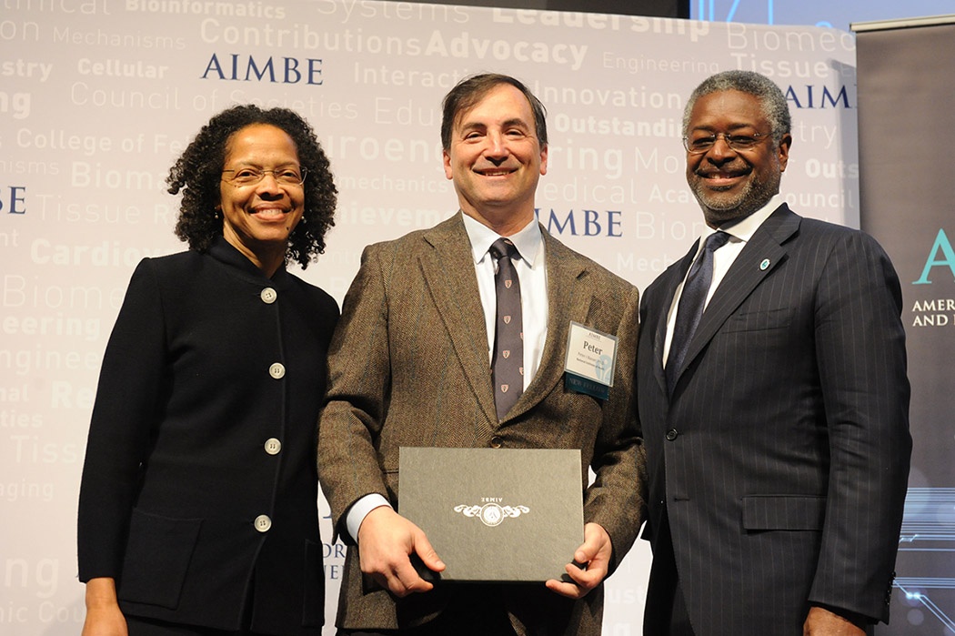
Dr. Peter Basser is awarded the Victor M. Haughton Award at the October annual meeting of the American Society of Functional Neuroradiology in Portland, OR. (NIH Record, November 17, 2017)
Dr. Ruiliang Bai received the Summa Cum Laude Merit Award at ISMRM 2017 (PDF 147 KB)
Dr. Ruiliang Bai received two Summa Cum Laude awards for outstanding posters at ISMRM 2016 (PDF 133 KB)
Dr. Ruiliang Bai received the Young Investigator Award at the 2016 Overseas Chinese Society for Magnetic Resonance in Medicine (PDF alternative [PDF 11 MB])
Dr. Dan Benjamini received the 2016 Giulio Cesare Borgia Prize at MRPM13
Dr. Basser et al. was honored as the author of one of ISMRM's "30 most influential MRM papers" at ISMRM 2014 in Milan, Italy. Photos here: https://twitter.com/alex_leemans/status/466644304342306816
The 30 most influential MRM papers that shaped the field of MRI! Unique event to meet the pioneers! #ismrm14 @ISMRM pic.twitter.com/2VVIFlxNtK
— Alexander Leemans (@Alex_Leemans) May 14, 2014
Drs. Peter Basser, Denis LeBihan, and Carlo Pierpaoli receive the Award for Excellence in Technology Transfer at the 2013 FLC Mid-Atlantic Regional Meeting in Leesburg, VA. (PDF alternative [PDF 67 KB])
Peter Basser, 2010 ISMRM Fellow
- NICHD’s Basser Honored for MRI Technology. (2010). NIH Record, 62(15), p.5.
Dr. Basser receives the 2008 ISMRM Gold Medal
Carlo Pierpaoli, 2008 ISMRM Fellow
Outreach
Click here for info about the NIH Conference, "Diffusion Tensor MRI: From Bench to Bedside"
IP
Smith, C. (2002) NIH commercializes new imaging technique. Nature Medicine 8(9): 906.
NIH Backgrounder, "Diffusion Tensor Magnetic Resonance Imaging"
Software
Grants
2019—SQITS lab member Nathan Williamson was awarded three years of NIH funding through a Postdoctoral Research Associate Training (PRAT) fellowship to study new methods to measure water motion in living organisms with magnetic resonance.
USUHS Center for Neuroscience and Regenerative Medicine grant: "Development of Bench and Preclinical MRI Methods to Assess Glymphatic Clearance in the Living Brain"
US-Israel Binational Science Foundation grants
2018 Bench-to-Bedside Award, funded by Intramural Research Program (Director's Challenge Award): "Localization of Epileptogenic Foci using Diffusion Weighted MRI"
NIH BRAIN Initiative funded awards
 BACK TO TOP