 The mission of the Developmental Translational Imaging Affinity Group is to understand the biology of normal/abnormal development primarily by the application of innovative, state-of-the-art cellular, molecular, and organ imaging methods. This line of research is intended, inter alia, to provide new insights into the basis and underlying causes of chronic diseases and disorders and to monitor and assess their sequelae.
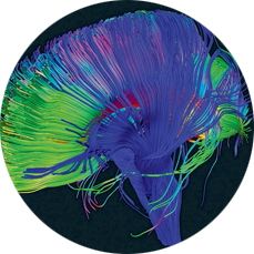
Quantitative Imaging and Tissue Sciences (Basser) invents, develops, and translates novel in vivo microstructural and functional MRI methods designed to measure salient properties of the developing brain and assess and characterize their changes in various diseases and disorders. These novel quantitative imaging biomarkers are also used in neuroscience applications to characterize brain network connectivity and dynamics, as well as in the clinic, to assess brain tissue microstructure and architectural organization. Significant efforts are being expended to develop imaging applications of portable, low-cost, low-field MRI scanners, such as the diagnosis of traumatic brain injury.
Translational Biophotonics (Gandjbakhche) uses multi-disciplinary approaches to devise “point-of-care” functional imaging technologies and methodologies that translate benchtop studies to the bedside. For example, near infrared spectroscopy and electroencephalography are used to assess biomarkers for a wide range of brain developmental abnormalities and injuries, specifically, but not limited to, cognitive and behavioral disorders in children and traumatic brain injury. The laboratory explores endogenous (scattering and absorption) and exogenous (using fluorescence probes) optical contrast mechanisms for characterizing abnormal development and function in tissues and organs, such as the placenta. They also are involved in clinical and preclinical studies aimed at characterizing growth and development of various abnormal tissues and monitoring the efficacy of their treatment using photonics methods, such as fluorescence lifetime and multi spectral imaging.
Biomedical Optics (Tromberg) develops models, methods, and devices for understanding and controlling light interactions with biological tissues. These methods are used to perform real-time quantitative measurements of clinically-relevant information, including tissue blood flow, oxygen extraction, metabolic rate of oxygen consumption, and body/tissue composition (lean mass, hydration, and fat mass). Advanced capabilities include continuous dynamic monitoring of intrinsic physiological signals that can be used in feedback optimization for guiding therapies and clinical decision making. Our technology development effort includes portable, bedside, non-contact and wearable sensor platforms, as well as the design and integration of probes into instruments for minimally invasive surgical feedback and guidance.
Pregnancy Research (Romero) investigates the biology of normal pregnancy and its most frequent complications such as premature labor, fetal death, preeclampsia, maternal and fetal infections (clinical chorioamnionitis), fetal growth disorders, and congenital anomalies--conditions which account for the excessive rate of infant and maternal mortality in the United States. The Branch conducts clinical and translational research and develops methods to improve the diagnosis, treatment, prediction and prevention of pregnancy complications and reduce adverse pregnancy outcomes. The Branch has participated in the care of over 25,000 pregnant women and its contributions have improved the understanding of pregnancy and its complications. The Branch places a strong emphasis on the study of human parturition and the role of infection and inflammation in causing obstetrical complications.
Labs within this group:
 BACK TO TOP