RNAscope Assay
RNAscope assay is a novel proprietary technology for RNA in situ hybridization (ISH) that was developed by Advanced Cell Diagnostics, Inc. (ACD Bio). The main difference of RNAscope with standard RNA ISH is the use of a double-Z probe design strategy: target-specific probes (ZZ pairs) minimize nonspecific off-target noise signals. This in turn allows highly specific staining capable of single-molecule detection in cultivated cells or tissues (1). RNAscope assay is used in a wide range of applications: gene expression, point mutation, antibody, vaccine or other therapeutic agents’ validation, etc…(2,3,4).
RNAscope assay can be used different kind of tissues:
- Formalin-fixed paraffin-embedded tissue: due to high auto-fluorescence, paraffin sections chromogenic assays may be preferred.
- Fresh-frozen tissues: offers highest reactivity
- Perfusion fixed frozen tissues: for tissues with high blood content (heart)
Three RNAscope assay kits developed by ACD Bio are widely used in biomedical research:
- RNAscope 2.5 HD Assay: Chromogen-based assay that produces sensitive and reliable result. Ideal for projects focusing on expression of one (possibly two) specific genes. The sample will be imaged in transmission mode.
- RNAscope Fluorescent Multipex Assay: fluorescence-based assay that allows simultaneous detection of up to 5 genes in complex tissues such as brain and tumors. If imaging on the MIC slide scanner, the sample must be stained with DAPI for tissue detection and focusing, thereby limiting the number of genes to 4 (green, red, far-red and infra-red fluorescence channels).
- RNAscope HiPlex Assay: Powerful fluorescence-based assay that allows detection of up to 24 genes in complex tissues (24 markers plus DAPI on the MIC slide scanner).
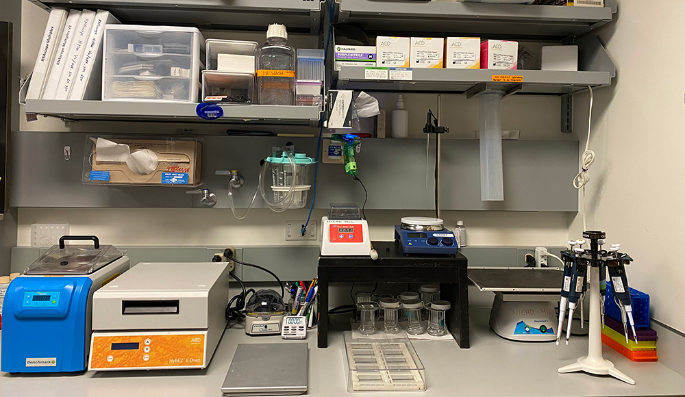
The MIC is fully equipped to handle RNAscope assays on fresh-frozen or perfusion-fixed frozen tissue. A fully equipped RNAscope bench that includes an ACD Bio HybEZ II Oven is available. Samples that do not use the infra-red (Cy7) channel of the ACD kit can be imaged on any microscope. Sample with the Cy7 channel must be imaged on either the Zeiss Axioscan automated slide scanner or the Zeiss wide-field microscope.
Note the MIC will not process large batches of RNAscope samples but rather train you on how to perform the staining. Contact Dr. Ling Yi (yil@mail.nih.gov) for details.
 BACK TO TOP