Histology
The sample preparation branch of the MIC is designed to take users through the steps of preparing high-quality samples for microscopy: dissection, fixation, cutting, immunostaining and mounting on slides. We also provide sample troubleshooting and technical advice on protocol optimization, antibody selection, and mounting media to help users obtain the best possible sample for imaging.
Our mode of operation is based on providing training and resources. We do not have the staff to undertake large histology workloads. Investigators who need to outsource large projects are encouraged to contact either one of the intramural histology facilities, or an outside provider such as Histoserv .
Available equipment includes:
- Gravity-driven small animal perfusion on a down-draft system (animal protocol required).
- Dissection platforms.
- Reagents and supplies for cryoprotection and cryo-embedding.
- ACDBio RNAScope ovens (3x)
- Sectioning equipment: Leica VT1000S Vibratome (50 – 1000 µm floating sections, fresh or fixed); Leica SM2010R Sliding Microtome (20 – 60 µm floating sections, fixed only); Leica CM-3050S Cryostat (5 – 100 µm frozen sections; fresh or fixed; thin sections on glass; sections >25µm can be floated); Brain Matrices (0.5 and 1 mm crude mouse or rat brain sections)
- Leica MZ6 Mobile Dissection Scope with LED Illumination.
- Bench space for sample processing, biosafety cabinet and 37°C incubators.
Pending availability of additional space, the MIC will provide paraffin-embedding services with a Leica Histocore Pegasus tissue processor and Biocut rotary microtome. Note we will NOT provide mass processing of large sample sets. Users will be trained on the technique and equipment, and will take over volume execution (Projected Feb. 2024).
The cryostat, RNAscope ovens, sliding microtome, perfusion fixation and paraffin processing can be reserved on our calendar .
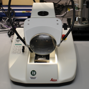
Leica Vibratome
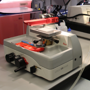
Leica Sliding Microtome
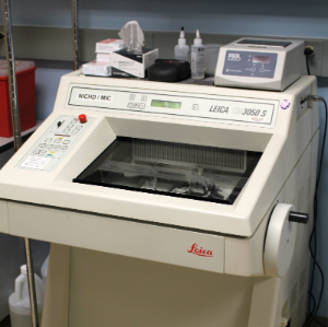
Leica Cryostat
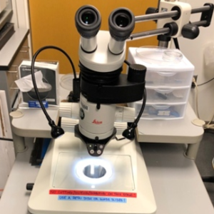
Dissection Microscope
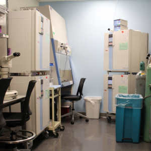
Tissue Culture Facilities
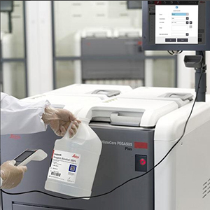
Paraffin processing
 BACK TO TOP