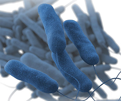
Three-dimensional computer-generated image of a group of Legionella pneumophila bacteria.
Credit: CDC/Sarah Bailey Cutchin
The bacterium Legionella pneumophila causes Legionnaires’ disease, a serious form of pneumonia. After entering the lung, L. pneumophila infects specialized immune cells called macrophages. Within each macrophage, the bacterium establishes small membrane-enclosed sacs, known as Legionella-containing vacuoles (LCVs), in which it multiplies before killing the host cell and infecting neighboring cells. Understanding how this process works promises to inform development of treatment and prevention strategies for Legionnaires' disease and related illnesses.
A recent study by the Machner Lab focused on how L. pneumophila controls expansion of LCVs to accommodate growing numbers of bacterial offspring. They found that the bacterium controls LCV expansion by fine-tuning the generation of molecules called lysophospholipids in the vacuolar membrane. Specifically, the bacterial enzyme VpdC converts the phospholipids that comprise the membrane into lysophospholipids. At moderate levels, lysophospholipids promote membrane fusion, for example, by allowing host cell components to integrate into the LCV to enhance the vacuole’s capacity.
Notably, elevated levels of lysophospholipids obstruct membrane fusion. The researchers found that overproduction of the VpdC enzyme blocked expansion of LCVs, trapping bacteria in the vacuoles and reducing their ability to proliferate.
The results provide insights into the mechanisms by which L. pneumophila regulates vacuole expansion. They also suggest that other pathogens may use similar strategies to exploit the features of membrane phospholipids for their advantage.
Learn more about the Cell and Structural Biology Group: https://www.nichd.nih.gov/about/org/dir/affinity-groups/CSB
 BACK TO TOP