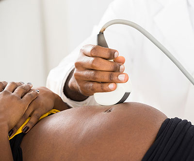
Uterine fibroids are the most common noncancerous tumors in women of reproductive age. Although many women with fibroids have uncomplicated pregnancies, some studies have suggested that having fibroids may raise the risk for abnormal positioning of the fetus, preterm birth, and cesarean delivery. Because of these potential links to pregnancy complications, scientists are working to better understand the changes that fibroids may undergo during pregnancy.
Researchers led by Katherine Grantz, M.D., from the Epidemiology Branch analyzed data from more than 2,700 participants in the Fetal Growth Studies-Singletons. Women in the study had up to six ultrasounds between weeks 10 and 41 of pregnancy. At each visit, sonographers recorded the number of fibroids and the volume of the three largest fibroids. Almost 10% of this racially and ethnically diverse group of study participants had fibroids visible on ultrasound during pregnancy.
On average, the total volume and number of fibroids declined from the first trimester of pregnancy to delivery. However, substantial variations emerged when researchers considered fibroid volume at the first ultrasound. On average, women with small starting fibroid volumes experienced increases in total volume, those with medium starting volumes saw almost no change, and those with large starting volumes experienced decreases. Volume changes also varied according to certain maternal characteristics. For example, non-Hispanic Black women, younger women, and women who were pregnant for the first time were more likely to experience fibroid volume decreases during pregnancy.
The findings expand knowledge of fibroid changes during pregnancy and may help clinicians counsel pregnant patients who have concerns about their fibroids and how they may change throughout pregnancy.
Learn more about the Division of Population Health Research (DiPHR): https://www.nichd.nih.gov/about/org/dir/dph
 BACK TO TOP