Signs that a placenta may malfunction later in pregnancy are often present in the first trimester and detectable by ultrasound. However, proper analysis of ultrasound images is difficult, time-consuming, and prone to human error. Researchers supported by NICHD through the Human Placenta Project are developing state-of-the art ultrasound screening tools that reliably measure the size and shape of the placenta and assess its blood supply early in a pregnancy.
Predicting Fetal Growth Restriction
Fetal growth restriction (FGR) is a condition in which the fetus fails to grow normally. Healthcare providers attempt to track fetal growth by measuring the pregnant woman’s abdomen, but this is a crude method at best. Identifying the risk for FGR early in pregnancy is important because of the serious risks during pregnancy, including of stillbirth, and later risks to a baby born too small, including increased risks of heart disease, stroke, and diabetes. Earlier diagnosis can lead to better outcomes.
Screening for Placental Insufficiency
FGR is often caused by placental insufficiency. It happens when the placenta does not develop properly or is damaged. One way to predict placental insufficiency is to measure its size early in pregnancy. Research shows that measurements of the placenta using ultrasound made by human operators are prone to error and may have low reproducibility rates. The Oxford researchers decided to investigate whether artificial intelligence could be used to train computers to measure placental volume using 3D ultrasound images. The automated method they created produced state-of-the-art results. The ultimate goal is to combine the automated measurements with other risk factors to predict placenta insufficiency, potentially providing a gold standard of care for FGR prediction during pregnancy.
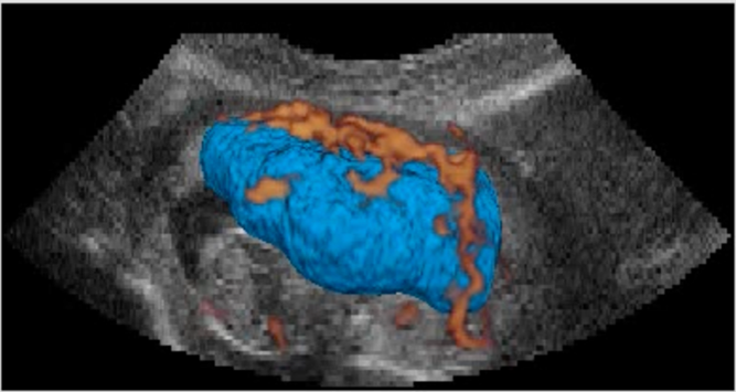
Caption: First-trimester placenta segmented with the AI tool, OxNNet
Credit: Sally Collins, M.D., Ph.D.
Detecting Placenta Adhesion
Placenta accreta spectrum (PAS) disorder is another potentially life-threatening condition for which better tests are needed. PAS results from placentas that do not detach from the uterus normally after birth, which can cause severe hemorrhaging for the pregnant woman. The ability to predict this condition would allow for proper high-risk care to be prescribed, saving lives. The Oxford group has used machine learning to teach a computer how to recognize abnormalities at the placenta-uterine interface.
New Gold Standard of Care
NICHD-supported researchers are on the verge of introducing a new gold standard of care by applying the latest in computer science to the analysis of ultrasound images. Studies are under way to validate their findings and show that the tool will, when combined with detection of other risk factors, provide increased prediction of potentially life-threatening placental abnormalities.
Learn more about the team
Principal Investigators:
Learn more about the HPP-funded project:
Development of real-time, automated 3D ultrasound tools for the study of placental invasion and morphology
 BACK TO TOP