Impeded blood flow within the placenta has been linked to pregnancy-related hypertensive disorders, including preeclampsia. However, the physiological mechanisms regulating placental blood flow are not well understood. Researchers supported by NICHD through the Human Placenta Project are using novel magnetic resonance imaging (MRI) techniques to study blood flow, oxygenation, and other physiological characteristics of the placenta—and their potential relationship to preeclampsia and other high-risk pregnancy conditions.
Understanding Placental Blood Flow Patterns in Pregnancy
Preeclampsia has been linked to poor blood vessel development and blood flow within the placenta. The condition can lead to poor regulation of blood pressure in the pregnant woman. Proper blood flow is essential for ensuring the fetus receives sufficient oxygen and nutrients during development.
To better understand blood flow and oxygenation patterns within the placenta, researchers from the Sir Peter Mansfield Imaging Centre in Nottingham, United Kingdom, used phase contrast angiography (PCA) and diffusion-weighted imaging (DWI) MRI techniques to compare these patterns in women with healthy pregnancies and in women with preeclampsia.
In one study, researchers found that in healthy pregnancies, blood flow speed decreased from the uterine wall through to the placenta. They also found high oxygen rates within the placenta, supporting previous hypotheses that slower flow is a more efficient way to transfer oxygen between mother and fetus. By contrast, in pregnant women with preeclampsia, researchers observed faster rates of blood flow and lower and more variable oxygen rates across the placenta compared with the healthy pregnancy group.
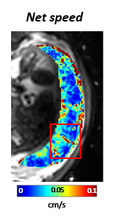
Caption: Map of the net speed of blood flow in the placenta at the level of the MRI voxels.
Credit: Penny Gowland
Identifying That the Placenta and Uterus Contract Independently
The same research team also identified a new physiological phenomenon, the “utero-placental pump,” by which the placenta and underlying uterine wall contract independently of the rest of the uterus to expel maternal blood. Researchers said this is an important discovery that provides a better understanding of the physiological mechanisms necessary for maintaining optimal blood and oxygen exchange levels between the mother and fetus.
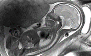
Caption: An MRI image of a fetus at 3T.
Credit: Penny Gowland
Initial Research Implications
Providing new insights into placental blood flow, oxygen transport, and how the placenta and uterus contract independently can help researchers develop new clinical tests to identify at-risk pregnancies. Such insights can also help guide clinicians in choosing the best management approaches for complicated pregnancies and assist in the early diagnosis of pregnancy-related conditions, such as preeclampsia. Additional research is needed to investigate the links between oxygenation and blood flow during contractions and identify what triggers these contractions.
Learn more about the team
Principal Investigators:
Learn more about the HPP-funded project:
Structure and function of the placenta from implantation to delivery: a next generation MRI approach
 BACK TO TOP