Despite its critical role in human development, the basic biology of the placenta is poorly understood. Novel magnetic resonance imaging (MRI) and other noninvasive imaging technologies are helping researchers reveal how the placenta functions during uncomplicated pregnancies and in ones where placental abnormalities exist. An international team of scientists supported by NICHD through the Human Placenta Project is using MRI and other imaging techniques to detail molecular transport and metabolism in pregnant mice, the placenta, and the developing fetus.
Novel Imaging Techniques
The team developed noninvasive MRI imaging techniques to study placental function in rodents. In an early study , the researchers showed they could characterize blood flow, transport, and oxygen exchange in the placenta of pregnant mice—eliminating the need to draw blood to complete these studies. In a later study , the team used new methods to compare changes in the placenta of normal mice with mouse models of fetal growth restriction (FGR) and preeclampsia. The team was able to characterize differences in placental development, setting the stage for clinical studies and potential diagnostic imaging in humans.
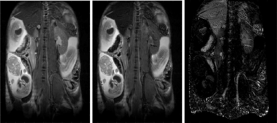
Caption: T fast spin–echo images of a pregnant mouse. Normoxic breathing gas (left); hyperoxic breathing gas (middle); difference image (right). Placental labyrinth, junctional zone, and myometrium can be clearly resolved in the image collected under hyperoxia.
Credit: Joel Garbow, Ph.D.
Mapping Placental Transport Mechanisms
The team also developed ways of mapping vascular microstructures and devised methods for detailing transport and metabolism of molecules important for normal fetal development. For example, they found that levels of biotin (vitamin H) were higher in the placenta than in maternal or fetal blood, and they also developed methods for mapping biotin transporter activity in the murine placenta. In the study , MRIs of pregnant mice were taken without exposing fetuses to contrast media, an important consideration for future human studies. The new method may also be used to study the transport of other molecules. Overall, noninvasive MRI studies in animal models set the stage for in vivo studies of human placenta function.
Learn more about the team
Principal Investigators:
Learn more about the HPP-funded project:
Integrated Placental Imaging: Novel Methods for Probing Function and Metabolism
 BACK TO TOP