A health care provider uses blood and urine tests that measure catecholamines and/or their metabolites to diagnose pheochromocytoma. Metabolites are biochemical substances that form when another substance is broken down in the body. Higher-than-typical amounts of these biochemical substances in the blood and/or urine can be an indication of the presence of a pheochromocytoma/paraganglioma.
Pheochromocytomas can secrete all, none, or any combination of catecholamines (epinephrine, norepinephrine, dopamine) and their metabolites, which are called metanephrines (metanephrine, normetanephrine, methoxytyramine). Multiple studies at the National Institutes of Health (NIH) have demonstrated the utility of measuring metanephrines in the blood for the diagnosis of pheochromocytoma/paraganglioma.1 This method is often recommended as the first diagnostic test when there is suspicion of a pheochromocytoma/paraganglioma.
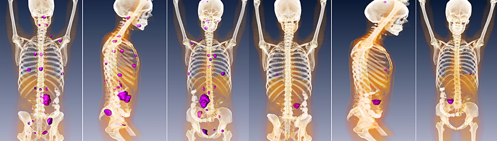
Tumors can also be found accidentally during nonrelated imaging studies. The location of a pheochromocytoma can be determined by using several imaging methods, including computed tomography (CT) and magnetic resonance imaging (MRI). CT scans use X-rays to produce detailed images of the inside of the body, while MRI uses magnetic waves to produce these pictures. A third imaging method that can be used to detect pheochromocytomas is MIBG (metaiodobenzylguanidine) scintigraphy. During this procedure, MIBG, a compound containing a small amount of radioactivity, is injected into a vein and is picked up by pheochromocytoma cells but not healthy cells.1 The body is scanned with a scanner that detects the MIBG. Any MIBG that is seen with the scanner can indicate the presence of pheochromocytoma cells.
 BACK TO TOP