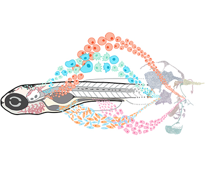
Illustration of the concept of single cell analysis during early zebrafish development.
Credit: J. Farrell, NICHD/NIH
Researchers rely on model organisms like zebrafish to identify how embryos mature from a single cell into distinct cell types, tissues, and organs. Many of the gene expression programs that direct embryonic growth are similar across fish and people, and findings in zebrafish can inform studies on human development and disease.
In a recent study from the Farrell Lab, researchers used a method called single-cell RNA sequencing to identify gene expression programs that are active in the first five days of zebrafish development. Collectively, this resource serves as a genetic atlas of early development.
- The atlas profiles nearly 490,000 cells from 62 timepoints during the first 120 hours after fertilization, with an average of 8,621 transcripts and 1,745 genes detected per cell.
- The study team then sorted these data into distinct cell types and cell states, identifying over 200 cell types present during this period of development.
- To highlight the atlas’ utility, the team focused on the development of understudied cells, including intestinal cells called BEST4+ cells, which are potentially linked to gastrointestinal diseases and cancer in people. Little is known about how these cells develop because they are absent in other common model organisms, such as mice.
- By using the atlas, the team computationally predicted the full developmental program of BEST4+ cells, including signals that initiate the cells’ development and transcription factors that carry out the process. These findings can be evaluated in model organisms or clinical samples to better understand the role of BEST4+ cells in human disease.
- The atlas, called Daniocell, is publicly accessible to the broader research community at https://daniocell.nichd.nih.gov. Additionally, microscopy images related to the study are also available.
NICHD co-authors of the paper include Abhinav Sur, Paulina Capar, Morgan Kathleen Prochaska, Gennady Margolin, and Jeffrey A. Farrell.
Learn more about the Cell Regulation and Development Affinity Group: https://www.nichd.nih.gov/about/org/dir/affinity-groups/CRD.
 BACK TO TOP