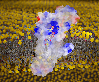
Visualization of fatty acyl CoA bound to membrane-embedded human DHHC20.
Credit: Jeremy Swan. The image was created using UCSF ChimeraX and Blender .
Thousands of proteins undergo a modification called S-acylation, in which fatty acids are attached to certain sites on the protein. This modification can be crucial for a protein’s proper function by altering its structure, interaction with other proteins, and other factors. S-acylation also has been implicated in diseases ranging from cancer to COVID-19, suggesting that developing ways to block the process could serve as potential treatment strategies.
In human cells, S-acylation is carried out by 23 integral membrane DHHC enzymes. DHHC enzymes transfer a fatty acid from a molecule called fatty acyl CoA to the target protein. Gaining a detailed understanding of this enzymatic reaction would help accelerate efforts to develop ways to block S-acylation.
The Banerjee Lab and their NIH colleagues used a combination of experimental techniques and computer simulations to visualize the earliest step in the S-acylation reaction—the recognition of fatty acyl CoA by a DHHC enzyme. They found that fatty acyl CoA binds to DHHC in a bivalent, or two-pronged, manner. Both the fatty acyl part of the molecule and the CoA part interact with different parts of the DHHC enzyme. Biochemical experiments revealed that both interactions are important for the enzymatic reaction to proceed.
These atomic-level snapshots of fatty acyl CoA bound to DHHC improve understanding of this critical early step in S-acylation. The techniques used in this paper could be applied to efforts to elucidate the entire enzymatic reaction.
Learn more about the Cell and Structural Biology Group: https://www.nichd.nih.gov/about/org/dir/affinity-groups/CSB
 BACK TO TOP