Placenta accreta spectrum disorders refer to a range of gestational pathologies in which the placenta invades the uterine wall, complicating childbirth. Early diagnosis during pregnancy can help reduce risks and improve outcomes associated with excessive maternal bleeding during delivery. However, current prenatal diagnostic tools often fail to detect these disorders, and some contrast agents have been linked to fetal safety issues. Researchers supported by NICHD through the Human Placenta Project (HPP) are developing and investigating new and potentially safer imaging contrast agents to facilitate earlier and more accurate detection of these potentially life-threatening conditions.
Diagnosing Patients at Risk
Rates of placenta accreta spectrum have increased in recent decades, and maternal death rates are higher for patients with the condition because severe hemorrhaging (bleeding) can occur during childbirth. Patients with placenta accreta spectrum are also more likely to require longer hospital stays and hysterectomy (surgical removal of the uterus).
Early and accurate detection of the condition during pregnancy is crucial for ensuring that these high-risk patients receive appropriate levels of maternal care. This includes access to medical staff trained in managing complex maternal and obstetric complications. However, current prenatal diagnostic imaging techniques, including ultrasound and magnetic resonance imaging (MRI), are not always able to detect the presence of the disorder and risk of severe complications. There are agents that improve image quality, but these "contrast agents" are associated with potential safety issues for the developing fetus.
Examining Use of Novel Contrast Agents for Improved Imaging
Researchers are developing and testing novel contrast agents that enable clearer imaging of placenta accreta spectrum disorders to facilitate early and more accurate diagnosis of the disorder. These novel agents, which do not cross the placenta, are also less likely to pose risks to the developing fetus.
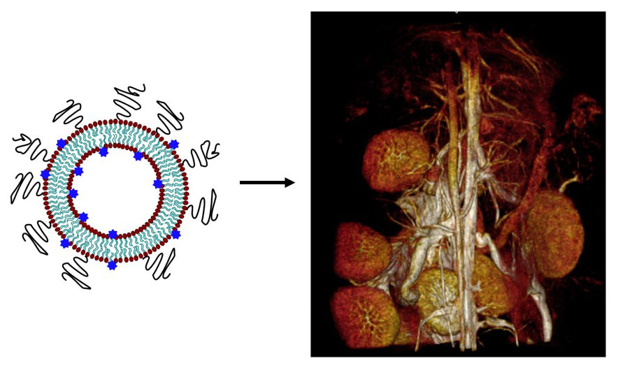
Credit: Translational Imaging Group (TIGr) at Texas Children’s Hospital and Baylor College of Medicine
Specifically, researchers are testing contrast-enhanced MRI using a novel liposomal gadolinium contrast agent (liposomal Gd). The gadolinium is caged within a very strong macrocyclic chelator, called DOTA, and conjugated to a phospholipid for presentation on the surface of the liposome bilayer. This approach leads to the generation of a very sensitive, high T1 relaxivity MRI contrast agent.
Furthermore, the nanoparticle formulation of Gd chelate prevents passive transport of Gd across the placenta and therefore protects the fetus against potential direct exposure to the agent. This allows for clearer visualization of placental margins and delineation of the retroplacental clear space. Clearer visualization of these and other structures during pregnancy and gestation can help identify placental abnormalities that are very difficult to observe using current techniques.
In one study, liposomal-Gd contrast agents enabled clearer visualization of the retroplacental clear space than conventional Gd chelate contrast agents did. In another analysis, contrast-enhanced MRI using liposomal-Gd contrast agent enabled visualization of the retroplacental clear space as early as day 12 of gestation in a pregnant mouse model (equivalent to mid-second trimester in humans), in addition to visualization of placental structural changes throughout the gestational period.
Initial Research Implications
Liposomal-Gd agents enabled visualization of the retroplacental clear space safely without crossing the placental barrier. Translating these agents to clinical use should dramatically improve the diagnosis and stratification of placenta accreta spectrum disorders, potentially enable molecular imaging of the placenta, and reduce fetal exposure risks associated with traditional small molecule contrast agents. NICHD researchers are planning to conduct a clinical study in the near future.
Learn more about the team
Principal Investigator(s):
Learn more about the HPP-funded project:
Molecular and vascular MRI of placenta accreta
 BACK TO TOP