Postdoctoral Position
Studying the meninges and the neurovascular interface in the zebrafish
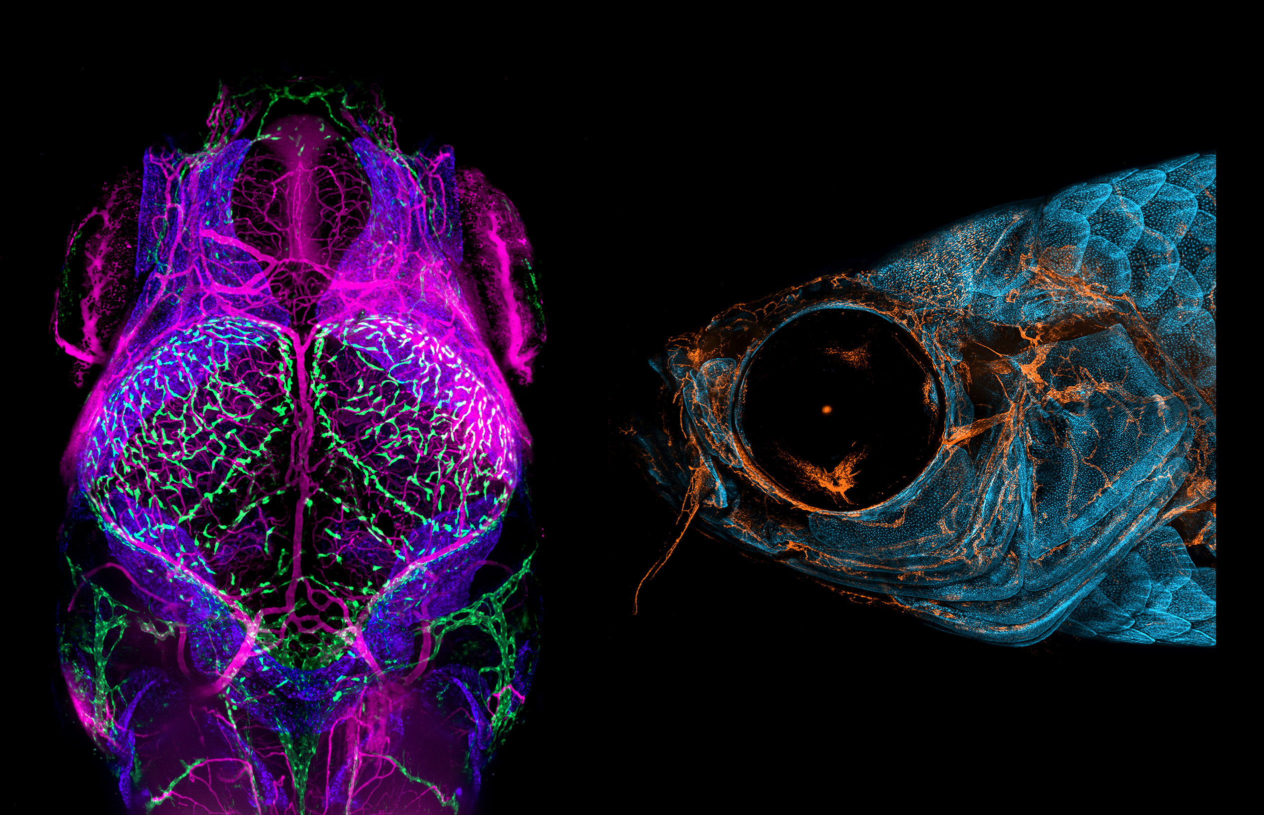
A fully funded postdoctoral position is available to study the developmental and function of the brain meninges and/or the neurovascular unit in the zebrafish in the laboratory of Dr. Brant Weinstein in the NICHD Division of Developmental Biology (DDB) in Bethesda, Maryland. The Weinstein laboratory uses a variety of cutting-edge molecular, cellular, genetic, transgenic, microscopic imaging, and next-gen sequencing approaches to study the meninges and meninges-associated cell types and the brain neurovascular interface. The meninges are complex, multilayered, highly vascularized tissues surrounding the brain that play a critical role in brain homeostasis and protection and are involved in a variety of brain pathologies. Our laboratory recently discovered that the zebrafish has a meningeal architecture very similar to that of mammals, and we are exploiting the advantages of the fish as a powerful new model for genetic and experimental dissection of this critical tissue. We are also interested in using the zebrafish to better understand the neurovascular interface throughout the brain, using the sophisticated genetic and imaging capabilities of the zebrafish.
The scientific environment, resources, and stipend support for this position are superb. Learn more about research in the Weinstein lab.
Interested applicants should have a Ph.D. or M.D. and less than 3 years' postdoctoral experience. To apply, send a curriculum vitae, bibliography, cover letter with a brief description of research experience and interests, and the names of 3 references (with phone numbers) via e-mail to weinsteb@mail.nih.gov and to amy.parkhurst@nih.gov.
The NIH is dedicated to building a diverse community in its training and employment programs.

Click image to enlarge.
 for more examples of vessel imaging in the meninges.
 BACK TO TOP