Characterization of Tissue Structures by Polarized Light
We develop novel methods to analyze tissue structures using polarization imaging. The specific aims include:
- Enhancement of biological tissue hidden structures by analyzing regions with statistical similarities with polarized light.
- Development of a handheld camera with liquid crystal retarder for active polarization imaging of the biological texture.
Enhancement of hidden structures of early skin fibrosis using polarization degree patterns and Pearson correlation analysis
Images of correlation coefficient corresponding to the same central point of ROI as in Fig. 2 For ROI dimensions of a 9x9 pixels and b 35x35 pixels
Many components of biological tissues, such as collagen, muscle fibers, keratin, retina, and glucose demonstrate polarization properties due to their anisotropy. As a result, depolarization of initially polarized light can be observed. This effect is strongly dependent on the bulk optical properties of the tissue (absorption and scattering coefficients) and optical anisotropy. Mapping the degree of polarization may carry valuable diagnostic information about the superficial and subsurface structures of the skin and other tissues, and may contain information that one cannot observe visually or photographically. A statistical analysis of data, involving a noise filtering procedure, can help to exclude distortions in the polarization degree maps due to image blurring and enhance observability of the hidden subsurface structures. As a result, characteristic scales and directionality of these structures can be quantified. One of potential applications of the suggested methodology is to characterize the state of the uterus to predict pre-term birth. According to the literature, cervical ripening is likely to result in pre-term changes in the uterus collagen structure. Polarization imaging can potentially be used to detect these changes.
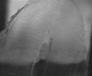
In order to investigate the anisotropy of mouse and human skin, we have devised an imaging system based on polarized videoreflectometry. Equi-intensity profiles of focused light, backscattered from the sample, are fitted with ellipses that appear to follow the orientation of the collagen fibers. A qualitative difference between intensity distributions for cross- and co-polarized orientations of the polarization analyzer is observed up to a distance of 1.5 to 2.5 mm from the entry point for the human skin. This method may be useful to assess skin fibrosis resulting from X-ray radiation treatment of cancer through the skin. We have shown that analyzing polarization degree images reveals the hidden structure in mouse skin of early fibrosis created by X-rays. The specific pattern of the surface distribution of the degree of polarization can be used to detect and analyze structures of initial skin fibrosis at the early stages of abnormality, when it is barely visible. We have devised a new data processing algorithm which involves Fourier filtering out of the high-frequency noise with subsequent Pearson correlation analysis. The latter provides information about characteristic scales of skin structures and their directionality. The technique of Pearson correlation coefficient analysis is used to reduce blurring of these features by unpolarized backscattered light and to visualize the regions of high statistical similarities within the noisy tissue images. We have shown that under certain conditions, such correlation coefficient maps are determined by the textural character of tissues and not by the chosen region of interest, thus, providing information on subsurface structural features of tissues from polarized light images.
Future Direction
Our hypothesis is that additional information, obtained from polarization images from the pre-term uterus can be used to predict cervical ripening and potentially diagnose pre-term birth. To verify our hypothesis, we intend to develop a handheld camera for active polarization imaging of biological tissue structures. The camera allows illumination with linearly polarized light and consequently captures two orthogonally polarized images using a liquid crystal retarder and polarizer. It will take approximately 150 ms to capture these images. No rotation of the optical elements is necessary. The camera is automated and data processing is realized by our software algorithm, which produces a real-time analysis of hidden features of biological tissues by Pearson analysis of cross-polarized and polarization degree images. The one-axis long working distance (300 mm) microscope with object illumination by collimated linearly polarized beam of intensive 850 nm wavelength LED is to be designed. A short length Galilean telescope will be used to provide a high quality magnified image at the plane of CCD matrix. We plan to incorporate a liquid crystal retarder and specially developed software into the commercial colposcope, creating the prototype of the instrument. The preliminary phantom experiments demonstrate the ability of the designed system to visualize the collagen network at ~10-20 mm scales. In collaboration with medical doctors we plan to analyze the results of in vivo tests in order to optimize potential informational/diagnostic content of the data.
 BACK TO TOP