Instrumentation
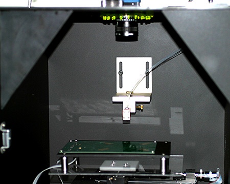
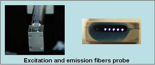
The in vivo optical imaging system for small animals involves scanning a single source-multiple detectors array over the surface of an object. Unlike commercially available imaging systems, the light source and detection fibers were placed at known distances from the source. Using several separations between the source and detectors allows probing more effectively at different depths of the medium. The imager scanned in a raster pattern over skin or other tissue surfaces to produce a real-time two-dimensional image. A cooled, charge-coupled device (CCD) camera is also used to guide the scan to the region of interest (ROI) and to measure the fluorescence intensity distribution, which helped to locate the tumor inside the tissue. Fluorescence lifetime can vary in response to changes in the immediate environment such as temperature, pH, etc.
 BACK TO TOP