Research
We are a small multidisciplinary team that uses a combination of cellular, molecular, genetic, biomechanical, live imaging and computational approaches to understand the development of the posterior lateral line primordium. This includes the use state-of-the-art microscopy, image processing and the development of multi-scale computational models to understand the self-organization of cell-fate, morphogenesis and migration of the lateral line primordium.
Research Approaches
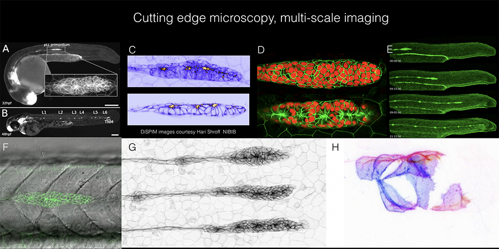
- Using multiscale imaging and image processing to visualize and characterize dynamics of morphogenesis and collective migration of the posterior lateral line primordium
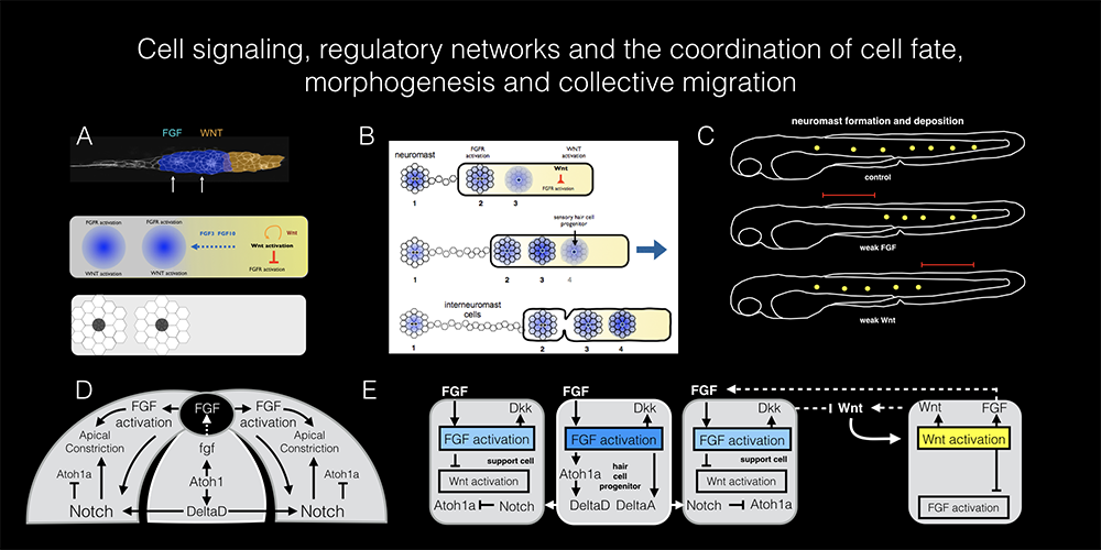
- Understanding cell signaling, regulatory networks and coordination of cell fate, morphogenesis and collective migration
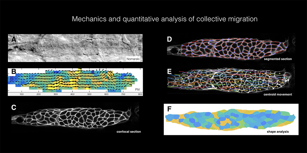
- Mechanics and quantitative analysis of collective migration
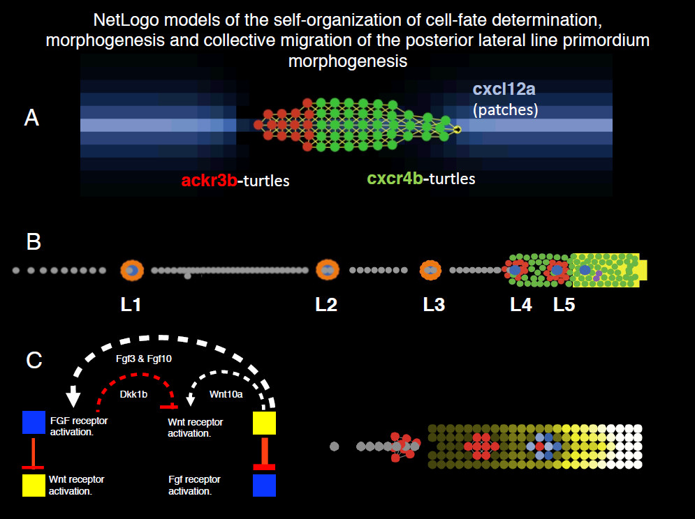
- Development of multiscale computational models of the self-organization of cell fate, morphogenesis, and collective migration of the posterior lateral line primordium
Click image to view.
Click image to view.
Click image to view.
Click image to view.
 .) B. A model that uses measured parameters including initial cell number, proliferation rate, the rate at which the Wnt system shrinks, and migration speed to predict the pattern of neuromast formation and deposition by the migrating primordium (Dalle Nogare and Chitnis 2017 ( https://doi.org/10.1016/j.mod.2017.04.005 ) C. A model that shows how mutually inhibitory interactions between Wnt and Fgf signaling meet theoretical requirements of reaction-diffusion patterning with local activation of Wnt coupled with its indirect long range inhibition system that can determine self-organization (Semin Cell Dev Biol. 2020 Apr;100:186-198. doi: 10.1016/j.semcdb.2019.12.015. Epub 2019 Dec 31).
 BACK TO TOP