A healthy, functioning placenta provides the necessary oxygen and nutrients to the growing fetus during pregnancy. Interdisciplinary research teams supported by NICHD through the Human Placenta Project (HPP) are studying new imaging techniques to more accurately identify and monitor placental dysfunction to predict high-risk pregnancies.
Using 3D Ultrasound and 4D Flow MRI to Monitor Placental Health
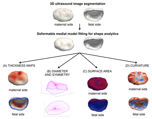
Placental volume is an indication of overall placenta size. Low placental volume (PV) early in pregnancy is linked to very low birth weight, placing infants at risk for developmental delays and neurocognitive disorders. In one study, the research team developed a new method for fully automating the analysis of 3D ultrasound (3DUS) images to accurately calculate placental volume. This new method also significantly reduced the time needed to measure PV using 3DUS in a clinical practice setting.
Impeded blood flow within the placenta causes oxygen deprivation during fetal development, sometimes leading to neurocognitive conditions and congenital heart problems in the infant. In another study, the research team successfully tested 4D flow magnetic resonance imaging (MRI) to measure the pulse of the uterine artery. This enabled the team to detect placental blood flow problems linked to high-risk pregnancies.
Credit: Nadav Scwhartz, M.D.
Combining Diffuse Optical Spectroscopy and Ultrasound to Measure Placental Oxygen
Directly measuring blood oxygen levels in the placenta can provide important insights into placental function. However, this often requires using invasive techniques that are not practical for bedside use. The research team recently developed a novel noninvasive diagnostic tool combining diffuse optical spectroscopy and ultrasound and a new algorithm methodology to perform calculations. In a pilot study, researchers succeeded in using the novel device and methodology to noninvasively measure placental oxygen levels deep (up to 4.2 centimeters) below the body surface, underneath intervening tissue layers. Moreover, the device helped investigators predict adverse pregnancy outcomes, as well as maternal vascular malperfusion (a type of placental injury caused by abnormal blood flow), by looking at how these novel measurements changed during maternal hyperoxygenation.
Initial Research Implications
Emerging technologies to identify placental dysfunction early in pregnancy can predict complications and potential for adverse outcomes. Automating the analysis of 3DUS images and integrating the use of 4D flow MRI can potentially serve as a gold standard for practical diagnostic tools. Noninvasive monitoring of placental oxygenation using diffuse optical spectroscopy and ultrasound can provide real-time bedside readings of placental oxygen levels. This can help researchers continuously collect placental data for use in future studies and help bedside clinicians with the early detection of placenta-related adverse pregnancy outcomes.
Learn more about the team
Principal Investigator(s):
Learn more about the HPP-funded project:
Developing a multi-modality, paradigm-shifting approach for in vivo assessment of the human placenta and the impact of maternal nutrition on its development and function
 BACK TO TOP