
New model system promises to spur research into developmental health and disease
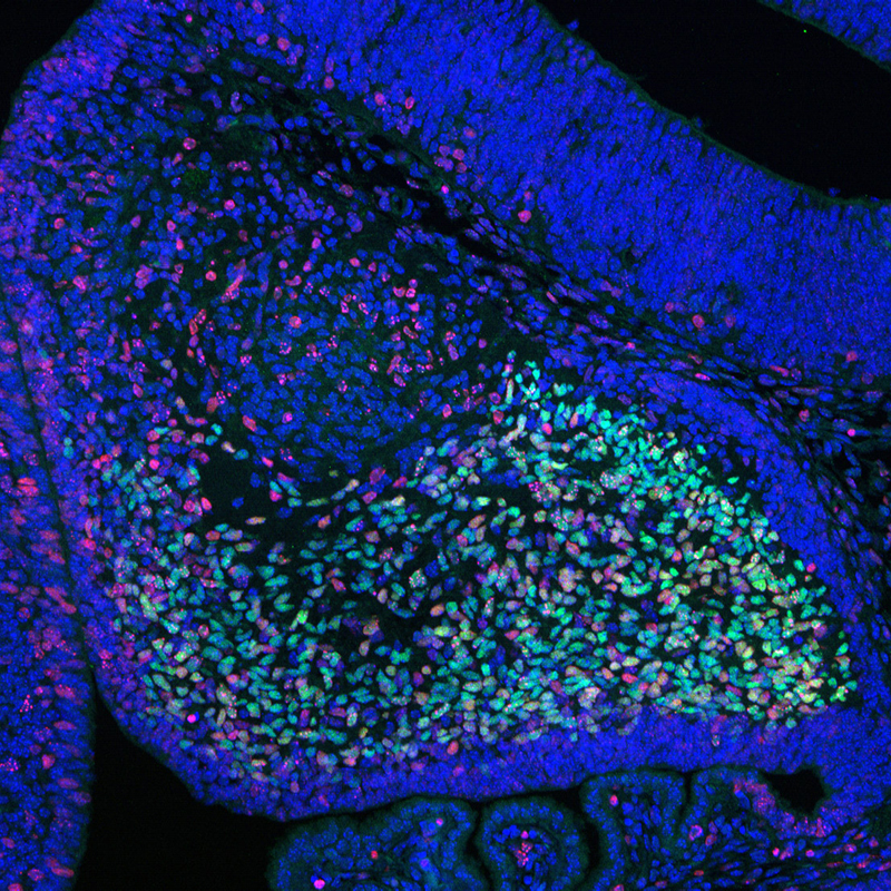
Credit: Vittorio Sartorelli, M.D., Laboratory of Muscle Stem Cells and Gene Regulation, National Institute of Arthritis and Musculoskeletal and Skin Diseases, NIH
WHAT:
Scientists funded in part by the National Institutes of Health have generated a mouse embryo model, or embryoid, that develops beyond neurulation—formation of the neural tube, which gives rise to the central nervous system—and closely mirrors natural mouse embryos 8.5 days after fertilization. These mouse embryoids offer a promising model system for research into factors affecting mammalian embryonic development and disease. The work is led by Magdalena Zernicka-Goetz, Ph.D., of the California Institute of Technology, Pasadena, and the University of Cambridge, U.K., and is partly supported by NIH’s Eunice Kennedy Shriver National Institute of Child Health and Human Development.
To assemble the mouse embryoids, the researchers combined mouse embryonic stem cells (ESCs) with two types of mouse extraembryonic stem cells that give rise to the yolk sac and placenta. This combination enabled development of all brain regions, in contrast to embryo models that are derived from ESCs alone and lack extraembryonic tissue. The resulting mouse embryoids also developed a neural tube flanked by somites—precursor cells that give rise to the vertebrae, skeletal muscle, cartilage and other structures. The mouse embryoids had a gut tube and precursor cells for formation of sperm. The whole structure developed within a yolk sac that formed blood islands, structures in which blood cells are formed to support the embryo and contribute to the circulatory system.
Additionally, the neural tube of the mouse embryoids responded to a developmental challenge much like the neural tube of natural mouse embryos. The scientists assessed the effects of a lack of Pax6, a protein required for brain and eye development. The neural tubes of mouse embryoids assembled with mouse ESCs lacking Pax6 appeared identical to those that develop in natural mouse embryos lacking Pax6. The authors conclude that mouse embryoids can serve as an experimental model to dissect the genetic and epigenetic factors that regulate development and to screen for the effect of chemicals on mammalian embryo development.
WHO:
Diana W. Bianchi, M.D., NICHD Director, is available for comment.
To arrange an interview with Dr. Bianchi, please e-mail nichdpress@mail.nih.gov or call 301-496-5133.
REFERENCE:
Amadei A, Handford CE et al. Synthetic embryos complete gastrulation to neurulation and organogenesis. Nature DOI: 10.1038/s41586-022-05246-3 (2022).
###
About the Eunice Kennedy Shriver National Institute of Child Health and Human Development (NICHD): NICHD leads research and training to understand human development, improve reproductive health, enhance the lives of children and adolescents, and optimize abilities for all. For more information, visit https://www.nichd.nih.gov.
About the National Institutes of Health (NIH): NIH, the nation's medical research agency, includes 27 Institutes and Centers and is a component of the U.S. Department of Health and Human Services. NIH is the primary federal agency conducting and supporting basic, clinical, and translational medical research, and is investigating the causes, treatments, and cures for both common and rare diseases. For more information about NIH and its programs, visit https://www.nih.gov.
 BACK TO TOP