
Findings improve understanding of the varying effects of the disease and may inform new treatment approaches
The gynecologic condition endometriosis has several differences in gene expression in the uterine lining, according to a comprehensive analysis of nearly 1,000 tissue samples by researchers funded in part by the National Institutes of Health. The differences stem from variations in DNA methylation—the binding of compounds known as methyl groups to DNA. Resulting from environmental factors, DNA methylation influences gene activity without changing DNA sequences and alters the genes’ protein products. Study of methylation patterns can yield information on how environmental factors affect the expression of individual genes, which may affect entire genetic networks and influence health and disease. The findings inform the search for the causes of endometriosis, shed light on why its effects vary among patients, and provide insights into developing new treatments for the disease.
The study was led by Linda Giudice, M.D., Ph.D., and Marina Sirota, Ph.D., of the University of California, San Francisco, and Stacey Missmer, Sc.D., of Michigan State University, working with colleagues at institutions in the United States, Europe, and Australia. It appears in Communications Biology. NIH funding was provided by the Eunice Kennedy Shriver National Institute of Child Health and Human Development (NICHD) and the National Cancer Institute.
Background
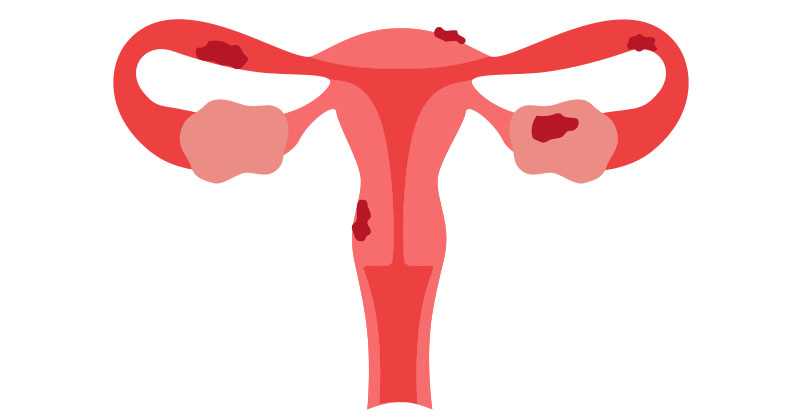
Endometriosis is a disease in which tissue resembling the lining of the uterus grows in other parts of the body, such as the ovaries, on the fallopian tubes, behind the uterus, on the bowels or on the bladder. Its causes are unknown, but it may affect anyone who menstruates, affecting roughly 10% around the world. It is associated with pain and infertility. Treatments involve surgery, hormone therapy, and pain-management approaches. Endometriosis is often classified into four stages (I-IV) according to the volume and type of endometriotic growths, their location, and other features.
To conduct the study, researchers analyzed samples of endometrial (uterine lining) tissue from 637 endometriosis patients (cases) and 347 participants without endometriosis (controls).
Results
DNA methylation in endometrial tissue varied according to the menstrual cycle phase. Differences in DNA methylation patterns accounted for 15.4% of the variation between cases and controls. The researchers believe this suggests that endometriosis may result from or promote multiple small methylation changes in the gene networks regulating endometrial function. The authors found the four most statistically significant differences in methylation patterns when comparing patients with stage III and IV endometriosis to controls.
The authors also found that 40% of variations in DNA sequences linked to genetic risk factors for endometriosis also were linked to differences in methylation patterns. These endometriosis-associated genetic effects on DNA methylation may lead to the identification of genes that play roles in the endometriosis disease process.
Significance
The authors believe their findings provide an important resource for understanding the genes involved in endometriosis and the development of therapies for it.
References
Mortlock, S., et al. Global endometrial DNA methylation analysis reveals insights into mQTL regulation and associated endometriosis disease risk and endometrial function. Communications Biology. 2023.
 BACK TO TOP