Findings could provide insight into early development, inform strategies for drug delivery
Credit: Eunice Kennedy Shriver National Institute of Child Health and Human Development, National Institutes of Health
Cells lining the umbilical vein secrete a newly discovered extracellular vesicle—a tiny, balloon-like structure that influences other cells—suggests a study by researchers at the National Institutes of Health. These extracellular vesicles, which the researchers termed multi-compartmented microvesicles, are released by cells lining the umbilical blood vessels. The tiny structures harbor and can release even tinier vesicles called exosomes that contain molecules known to interact with other cells and influence their function.
The findings may have implications for understanding fetal development and may inform strategies to use extracellular vesicles to deliver drugs and other therapies.
The study was conducted by Jennifer D. Petersen, Ph.D., Electron Microscopist, and Joshua Zimmerberg, M.D., Ph.D., Senior Investigator, at NIH’s Eunice Kennedy Shriver National Institute of Child Health and Human Development and colleagues. It appears in the Journal of Extracellular Biology.
Background
From bacteria to plants to animals, cells secrete extracellular vesicles containing proteins, enzymes, DNA, and other molecules capable of transmitting chemical messages between one cell and another. After they are released from a cell, extracellular vesicles can travel to other cells in the same vicinity or over longer distances via the blood, cerebrospinal fluid, or amniotic fluid. Recent studies have shown that extracellular vesicles may be used to detect certain types of cancer.
Endothelial cells line the inner surface of blood and lymphatic vessels. They control the exchange of oxygen and nutrients between the vessels and surrounding tissues. Endothelial cells release a large proportion of the extracellular vesicles found in the blood, which in turn release substances that regulate vessel functioning, prevent clots from forming on the endothelial cell lining, protect against inflammation, and promote new vessel formation.
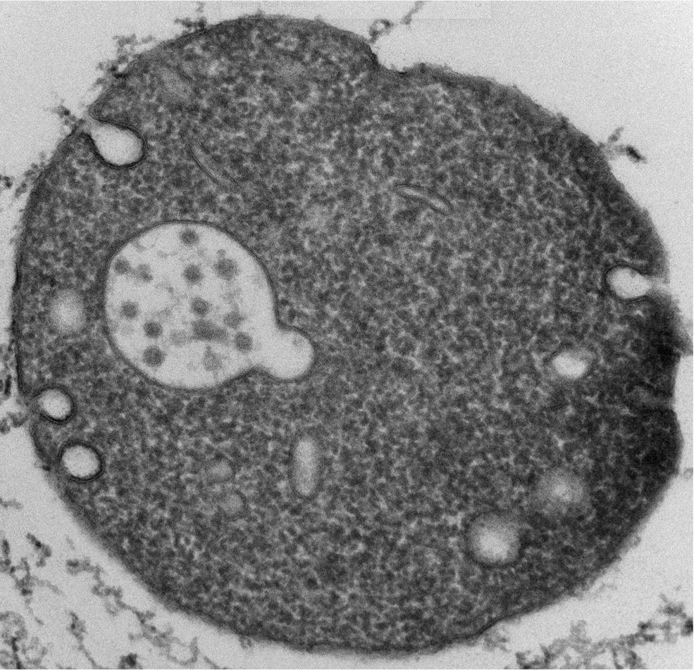
For the current study, researchers used electron microscopy to observe the formation of extracellular vesicles from cultures of endothelial cells that line the surface of the umbilical vein.
Results
The researchers first used umbilical vein endothelial cells to confirm the presence of finger-like protrusions on the cell surface from which extracellular vesicles previously have been shown to pinch off and enter the bloodstream. They observed that these protrusions often contain membrane-bound cellular organelles (structures within cells). They also observed vesicles outside the cells’ peripheries. These contained organelles similar to those seen in the protrusions, which the researchers believe indicates that they probably formed by pinching off from protrusions.
Because this new type of extracellular vesicle consisted of vesicles inside vesicles, the authors described them as multi-compartmented microvesicles (MCMVs). Some of the cellular organelles inside MCMVs included endoplasmic reticula and mitochondria, indicating that MCMVs may have their own source of metabolic energy.
Significance
The study identified multi-compartmented microvesicles, a new class of extracellular vesicle in umbilical cord vessels. Future studies are needed to determine the function of these vesicles, the molecules they release, and whether they can reach the vessels within the fetus and from there influence fetal development. Because the placenta is jointly maternal and fetal, they may represent a new means of communication between maternal and fetal cells.
Reference
Petersen, JD, et al. Endothelial cells release microvesicles that harbour multivesicular bodies and secrete exosomes. Journal of Extracellular Biology. 2023.
 BACK TO TOP