
Findings could inform efforts to treat osteoporosis and other bone loss disorders
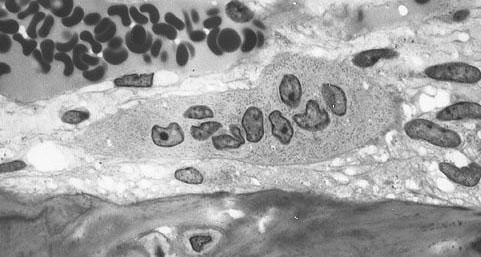
Credit: Public Domain, Wikimedia Commons, https://commons.wikimedia.org/wiki/File:Osteoclast.jpg

Researchers at the National Institutes of Health have determined that a protein interacting with RNA molecules in cell nuclei plays a role in forming osteoclasts—cells that break down old or damaged bone tissue so it can be replaced with new bone. The findings have implications for understanding degenerative bone disorders like osteoporosis and fibrous dysplasia.
The study was conducted by Jarred Whitlock, Ph.D., and other researchers in the laboratory of senior author Leonid V. Chernomordik, Ph.D., of the Eunice Kennedy Shriver National Institute of Child Health and Human Development (NICHD) and colleagues in the NICHD and the National Institute of Dental and Craniofacial Research, both at the NIH. Their study appears in Nature Communications.
Background
Osteoclasts are a type of bone cell that breaks down bone tissue. They work in balance with osteoblasts—cells that form new bone tissue to replace old tissue that osteoclasts have broken down. Osteoclasts have multiple cell nuclei. They are formed when several progenitor cells that originate in the bone marrow fuse together to form a large cell with multiple nuclei. The more fusions they complete, the larger osteoclasts become and the greater their ability to dissolve bone.
The number and size of osteoclasts are altered in many bone diseases and with the aging process. To gain insight into how the body controls osteoclast size and behavior, the study authors sought to identify the cellular mechanisms that control the fusion of osteoclasts.
Results
With cells isolated from blood donors, the researchers formed progenitor cells and then induced them to form osteoclasts. They discovered that a protein, referred to as La and thought to be present in cell nuclei, disappeared from the progenitor cells and then reappeared as they began to fuse into osteoclasts.
Using a variety of genetic, molecular, and cellular approaches, the researchers demonstrated that surface-bound La controls osteoclast fusion, regulating how large they become. The researchers tested whether blocking La’s surface function in osteoclasts could prevent the formation of excessively large osteoclasts in a disease, fibrous dysplasia. In a mouse model, they showed that blocking the compound resulted in fewer osteoclasts.
Significance
The authors believe that targeting La at the surface of osteoclasts may lead to treatments for bone disorders and age-related changes in osteoclast size and number, including osteoporosis and fibrous dysplasia.
Reference
Whitlock JM, et al. Cell surface-bound La protein regulates the cell fusion stage of osteoclastogenesis. Nature Communications. 2022. https://doi.org/10.1038/s41467-023-36168-x
 BACK TO TOP