
NIH-funded technique enables automatic detection of placental compartments, oxygen status and structural abnormalities
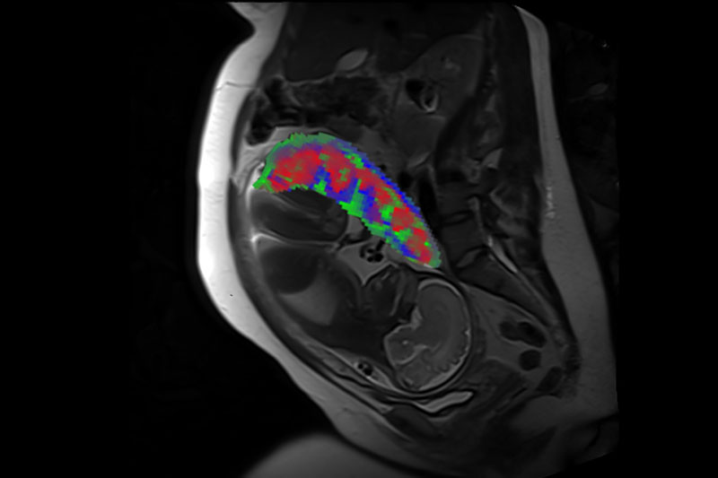
Credit: Wang Lab, Washington University in St. Louis
WHAT:
Researchers funded by the National Institutes of Health have developed a new method to process MRI scans to reveal the distinct compartments of the placenta, take measurements of oxygen levels in each region and determine if there are malformations in blood vessels (i.e., placental lesions). Obtaining this level of detail is currently not possible using standard MRI analysis methods. The small study was supported by NIH’s Human Placenta Project, which is led by the Eunice Kennedy Shriver National Institute of Child Health and Human Development (NICHD).
The study team, led by Yong Wang, Ph.D., at Washington University in St. Louis and Alison Cahill, M.D., at the University of Texas at Austin, developed analysis methods for MRI scans that are routinely collected in hospitals and healthcare facilities. Importantly, these types of MRI scans do not require contrast agents, which are only used during pregnancy for limited circumstances. The team’s machine learning method automatically processes MRI data to visualize separate placental compartments, including the intervillous space (the area where maternal blood enters to provide nutrients and gas exchange), placental vessels and placental tissue. Unlike current MRI analysis methods, which can only measure placental oxygen as an average across the entire organ, the new method can characterize oxygen levels within these discrete compartments.
The study team also used these region-specific measurements to look for differences between 22 study participants with healthy pregnancies and five participants who went on to develop pregnancy complications. While they did observe trends—highlighting the potential for this technique to detect specific pregnancy complications—further analysis is needed among larger groups of study participants.
Overall, the new method can serve as an objective, quantitative method to assess placental health during pregnancy. With additional validation and refinement of this technique, the method may be used by healthcare providers as a tool in the care of pregnant patients, especially those at risk for pregnancy complications. Early monitoring of the placenta can lead to better detection and prevention of pregnancy complications, including preterm birth, fetal growth disorders and preeclampsia.
WHO:
David Weinberg, Ph.D., Lead of the NICHD Human Placenta Project, is available for interviews.
To arrange an interview with Dr. Weinberg, please e-mail nichdpress@mail.nih.gov or call 301-496-5133.
REFERENCE:
Sun Z., et. al. Association of intraplacental oxygenation patterns on dual-contrast MRI with placental abnormality and fetal brain oxygenation. Ultrasound in Obstetrics & Gynecology DOI: 10.1002/uog.24959 (2023)
Journal cover image: https://obgyn.onlinelibrary.wiley.com/doi/abs/10.1002/uog.24939
###
About the Eunice Kennedy Shriver National Institute of Child Health and Human Development (NICHD): NICHD leads research and training to understand human development, improve reproductive health, enhance the lives of children and adolescents, and optimize abilities for all. For more information, visit https://www.nichd.nih.gov.
About the National Institutes of Health (NIH): NIH, the nation's medical research agency, includes 27 Institutes and Centers and is a component of the U.S. Department of Health and Human Services. NIH is the primary federal agency conducting and supporting basic, clinical, and translational medical research, and is investigating the causes, treatments, and cures for both common and rare diseases. For more information about NIH and its programs, visit https://www.nih.gov.
 BACK TO TOP