
Mouse study improves understanding of events leading to preterm labor and birth
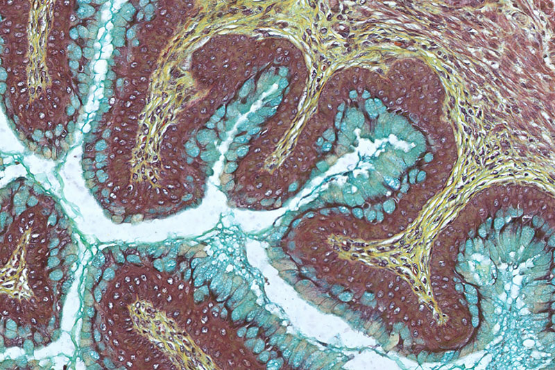
Credit: Pregnancy Research Branch, NICHD/NIH
NIH researchers have mapped out the cellular activity underlying infection-induced preterm labor in a mouse model. The findings, published in Cell Reports, provide a foundation for understanding how infection can prematurely trigger labor. With this knowledge, researchers can better identify potential ways to prevent a common cause of preterm birth in women.
Background
Preterm labor and birth occurs before 37 weeks of pregnancy. It is the most common cause of infant death and is the leading cause of long-term disabilities in children. Preterm labor and birth is often caused by infection of the amniotic fluid, also known as intra-amniotic infection. Researchers believe that the resulting inflammation triggers preterm labor, but the specific details of how this happens are still not fully understood.
Results
In the current study, researchers in the NICHD Pregnancy Research Branch and their colleagues used a technique called single-cell RNA-sequencing to generate a cellular map or atlas of the mouse uterus, decidua (specialized tissue that is considered the maternal-fetal interface), and cervix in a model of intra-amniotic, infection-induced preterm labor. Their work provides a detailed overview of the cellular composition and diversity of cells in these reproductive tissues.
The study team found that preterm labor dysregulated the activity of immune and non-immune cells, sometimes with similar activity across cells or tissues and sometimes in a tissue-specific manner. They also observed that certain cell types appeared to orchestrate communication in different tissues to induce labor. The study team compared these mouse observations to differences in human uterine tissues sampled during labor and saw similar trends. Such similarities reinforce the strength and applicability of their mouse atlas to human pregnancies.
Overall, the study findings highlight the complex cell-cell communication that takes place in the reproductive tissues and how such interactions are altered by the intra-amniotic inflammatory process of preterm labor.
Significance
The data from this study can serve as a reference for future research on preterm labor and birth. For example, the cell types and signaling pathways identified in this work can be validated in human studies. Once confirmed, additional research can target specific subsets of cells or signaling pathways in the reproductive tissues to potentially lower the risk of preterm birth.
Reference
Garcia-Flores V. et al., The single-cell atlas of the murine reproductive tissues during preterm labor. Cell Reports DOI: 10.1016/j.celrep.2022.111846 (2022)
 BACK TO TOP