Hospital staff typically use one of two methods for the hearing test. Both are quick (5 to 10 minutes) and safe.1
- Otoacoustic (pronounced oh-toe-uh-KOO-stik) emissions (OAE). This test determines if certain parts of the infant’s ear respond to sound. A miniature earphone and microphone are placed in the ear, and sounds are played. If the infant has normal hearing, the microphone picks up an echo reflected back into the ear canal. Failure to detect an echo means there may be a loss of hearing.
- Auditory brain stem response (ABR). This test evaluates the auditory brain stem—the part of the auditory nerve that carries sound from the ear to the brain—and the brain’s response to sound. Miniature earphones are placed in the ear, and sounds are played. Electrodes (small, sticky electric conductors) are placed on the infant’s head to detect the brain’s response to the sounds. If the infant’s brain does not respond consistently to the sounds, there may be a hearing problem.
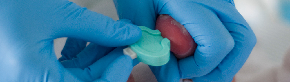
In some cases, hospital staff will perform pulse oximetry (pronounced ox-EM-i-tree) to measure how much oxygen is in the infant's blood. Pulse oximetry is usually performed at least 24 hours after birth. Hospital staff place a sensor on the infant's skin for a couple of minutes, and the sensor measures the level of oxygen in the blood through the skin.
Low blood oxygen can indicate that a newborn has heart problems. Pulse oximetry can help identify infants with a condition called critical congenital heart disease (CCHD).1 According to the Centers for Disease Control and Prevention, 25% of babies with congenital heart disease (one that is present at birth) have a CCHD.2 There is evidence that medical care is allowing more infants with CCHD to survive. One study found that 83% of infants with a CCHD who were born between 1994 and 2005 survived at least 1 year. Only 67% of infants born between 1979 and 1993 with a CCHD lived to 1 year.3
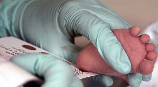
Some states require a second blood test that repeats the initial set of screenings.
- The first screening is performed 24 to 48 hours after the infant is born, ideally before the infant leaves the hospital. For some conditions, the screening is not valid if the blood is taken within 24 hours of birth.
- The second screening is performed when the infant is 10 days to 2 weeks old to ensure that the health care provider has the most accurate results possible.
 BACK TO TOP