
NIH scientists find drug’s active ingredient causes loss of key neurons
Fetal mice exposed in the uterus to delta-9-tetrahydrocannabinol (THC), marijuana’s primary psychoactive ingredient, lose a key subset of neurons, according to a new study by NICHD researchers. The findings offer an explanation on how marijuana abuse during pregnancy may lead to cognitive problems in children who are exposed to the drug in the womb. The study appears in the March 15, 2016, issue of Molecular Psychiatry.
Background
Marijuana refers to the dried leaves, flowers, stem, and seeds of the hemp plant, Cannabis sativa. It is the most commonly abused illicit drug in the United States, according to the Substance Abuse and Mental Health Services Administration. Although the Food and Drug Administration has not approved marijuana for medical use, several states have legalized the drug for this purpose.
Children born to women who used marijuana during pregnancy are more likely to have difficulty with problem-solving skills, memory, and remaining attentive. However, how the drug might cause these problems is not well understood.
According to researchers, THC binds to the cannabinoid family of brain receptors, overriding a normal brain communication circuit. During early brain development, signals from cannabinoid receptors are essential for the growth, migration, and specialization of neurons, which ultimately form the brain.
Results
The NICHD researchers focused their study on CCK-INTs, a neuron subtype likely susceptible to THC, because the cannabinoid receptor, CB1R, is found mainly on these neurons. The team, led by Chris J. McBain, Ph.D., from NICHD’s Program in Developmental Neurobiology, designed their study to investigate whether prenatal exposure to THC (or a synthetic mimic called WIN) affects the development and function of CCK-INTs in a mouse model.
The study team found that offspring exposed prenatally to THC or WIN had reduced numbers of CCK-INTs in the brain, particularly in an area called the hippocampus, which is involved in forming memory and regulating mood. CCK-INTs from drug-exposed offspring also had an altered shape, with fewer projections called dendrites. These effects were not seen in other neuron subtypes, suggesting a selective loss of CCK-INTs.
Importantly, the researchers determined that THC and WIN do not modify cannabinoid receptor signaling. Instead, these substances disrupt brain circuits by reducing the concentration of CCK-INTs. Furthermore, WIN-exposed offspring, compared to unexposed mice, spent significantly less time interacting with a new mouse—a change in social behavior. However, the study authors emphasized that much more research is needed to adequately characterize the role of CCK-INTs on behavior.
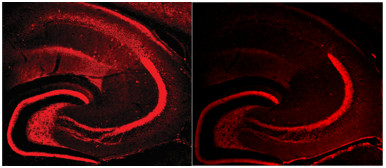
Credit: NICHD
Significance
As marijuana-derived products increase in prevalence across the country, researchers aim to determine the drug’s effects on the brain and on fetal development.
“Our findings are not just limited to understanding how prenatal marijuana exposure affects the developing brain,” said Dr. McBain. “The study also underscores the general importance of CB1R activity in neurodevelopment. It’s possible that an imbalance in these brain signals may play a role in disorders like autism, schizophrenia, and epilepsy.”
Reference
Vargish GA, Pelkey KA, Yuan X, Chittajallu R, Collins D, Fang C, and McBain CJ. Persistent inhibitory circuit defects and disrupted social behaviour following in utero exogenous cannabinoid exposure  . Molecular Psychiatry (2016)
 BACK TO TOP